 0
0
 0
0
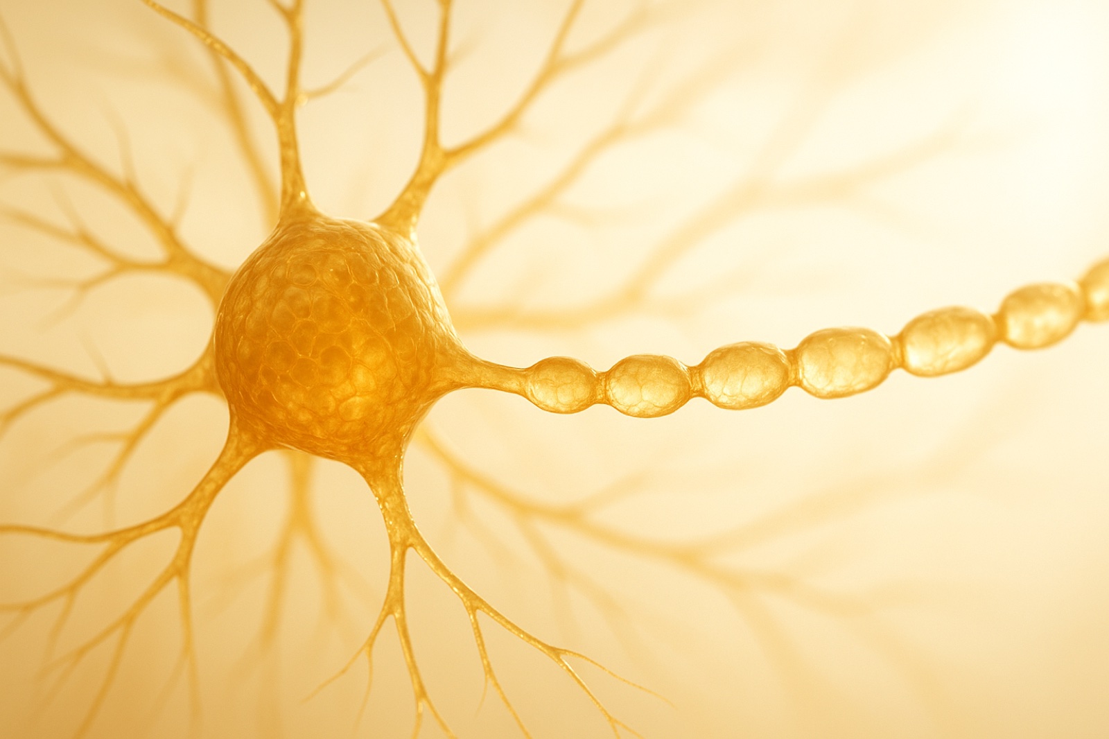
Open almost any biology textbook and you will find the same drawing. A brain cell with a round body, a spray of branches at one end, and one long smooth cable running out the other side. That cable is the axon. It is the wire. It is how one brain cell talks to the next.
So what do neurons look like, really? For about a hundred years the answer has been that smooth cable.
A team at Johns Hopkins Medicine says that picture may be wrong. In their images, the wire is not smooth at all. It looks like a string of tiny pearls — bulge, narrow, bulge, narrow, all the way down.
And a number of other neuroscientists have looked at those images and said: careful. You may have made the pearls yourself.
So what do neurons look like under a better microscope?
The Johns Hopkins work, led by Shigeki Watanabe with collaborators including Padmini Rangamani and Graham Knott, was published in the journal Nature Neuroscience on 2 December 2024. Most of it was done on mouse brain cells.
The difference was not a better lens. It was a different way of holding the tissue still.
For a century, the standard method has been chemical fixation: soak the tissue in chemicals, then dry it out, then look. The Hopkins team used high-pressure freezing instead — slamming the tissue to a frozen state so fast that the water inside never has time to form damaging crystals. The researchers compare it to freezing a grape instead of drying it into a raisin.
Frozen that way, the axons were beaded. The bulges sit at regular intervals and are not at the junctions where cells pass messages to each other, so they are not the swellings scientists already knew about. They are small enough that hundreds of them would fit across the width of a single human hair.
Then the team gave the cells an electrical jolt. The bulges swelled — on average about 8% longer and 17% wider — and stayed swollen for at least half an hour. The way signals travelled down the wire changed too, for at least an hour afterwards.
Here is the detail that is easy to read past: bigger pearls did not simply mean faster signals. The narrow connecting stretches between the bulges mattered as much as the bulges did. The shape of the gaps was part of the answer, not just the shape of the beads.
The part where other scientists push back
This is where the story stops being a tidy “textbooks were wrong” headline.
Christophe Leterrier, a neuroscientist at Aix-Marseille University, put the objection about as fairly as it can be put. He agrees the axon is not a perfect tube — but, in his words, “it’s not also just this kind of accordion that they show.”
Pietro De Camilli at Yale raised the sharper worry. Freezing is fast, he noted, but the sample still has to be handled to get there: “While quick freezing is an extremely rapid process, something may happen during the manipulation of the sample.”
Translation: the beads might not be how the axon lives. They might be how the axon reacts to being caught.
That objection is not unreasonable. Beaded axons are already a known sign of damage. When nerve fibres are injured or stressed, they bead up. So a picture of beaded axons is genuinely ambiguous. It might show normal anatomy nobody had seen properly. It might show a cell in distress.
Both sides are arguing about exactly the same thing
Notice what the disagreement actually is. It is not really about neurons.
The Hopkins team says the smooth tube is an artefact — that drying tissue out shrinks and smooths it, and a hundred years of textbooks have been drawing the consequence of a preparation method.
The critics say the pearls are an artefact — that something in the freezing or the handling creates the beads.
Each side is making the same accusation in opposite directions: the thing you are looking at was shaped by how you held it still.
And both are right that neither picture is a living brain, observed in place, going about its business. It is a specimen. Something had to be done to it before anyone could look. That is not a flaw in these particular scientists. It is the condition of looking at anything too small and too alive to sit still for the camera.
It is the same trap that kept a reef off the coast of Benin listed as dead for sixty years. The verdict came from dredges and sound equipment. Nobody had ever actually gone down and looked — and when someone finally did, the reef was alive.
What is actually settled, and what is not
Very little of this is finished, and it is worth being plain about that.
- Almost everyone now agrees the axon is not a perfectly smooth tube. How far from smooth is the open question.
- Follow-up work has looked at human brain tissue, published in the journal Neuron, but the samples came from epilepsy surgery and the study was mainly examining something else. It is a first look, not a settled answer.
- A separate team at the University of Edinburgh published work in PLOS Biology in July 2026 using live zebrafish to screen 880 compounds for effects on axon diameter, finding 33 that did something. That is a useful new tool, not a direct replication.
- Nobody yet knows how the pearling in a working axon relates to the beading seen in neurological disease. That question is wide open, and it is the one that matters most.
None of which makes the finding small. If it holds, the shape of the wire is not just packaging — it is part of how the signal moves, which means the brain may be adjusting its own wiring in a way nobody was watching for.
The quiet thing underneath
There is something worth sitting with here that has nothing to do with neurons.
Every picture we carry of anything came to us through some method of looking. A drawing. A measurement. A story someone told about a night they barely remember. Something was always done to the thing before we could see it, and that something leaves a mark on the picture.
Most of us walk around forgetting that. We do not hold our picture of a person, or of a stretch of our own life, as a preparation. We hold it as a photograph.
There is an old line of thought, far older than any microscope, that says human seeing is partial by nature — that we see now in fragments, as through dim glass, and that clarity is something that arrives later rather than something we already have. It was never meant as an insult. It was meant as relief. If the picture is partial, the picture can be revised. You are allowed to have been wrong about the shape of something for a hundred years and still be doing honest work.
That is a strangely freeing thing to know about yourself. It sits close to why certainty is so often the moment the checking quietly stops.
Where that leaves the textbook drawing
Probably in need of a redraw — though not the one the headlines promised.
The honest version of that picture is not a smooth tube and probably not a neat string of pearls either. It is something in between, still being argued over, drawn by people who keep telling you which method they used and what that method might have done.
Which is, when you think about it, the good version of this. A field that keeps arguing about its own preparation methods is a field that is still looking. It is the same reason a study about ageing brains is more interesting when it admits what it cannot say about brain shrinkage as when it announces what it can.
Somewhere behind your eyes, right now, a few hundred trillion of these connections are doing whatever it is they actually do. Nobody has ever seen one alive, in place, undisturbed. We are still working out what the wiring looks like — and we are only a hundred years into asking.
Questions people are asking
What do neurons look like?
A neuron has a cell body, branching extensions called dendrites that receive signals, and a single long fibre called an axon that sends them. Textbooks have drawn the axon as a smooth tube for around a century. Research published in Nature Neuroscience in December 2024 by a Johns Hopkins Medicine team found that in flash-frozen mouse tissue, axons instead appear as repeating bulges connected by narrow segments — a shape often described as pearls on a string. Other neuroscientists dispute whether that shape is how axons exist in the living brain.
Are axons smooth tubes or pearls on a string?
This is genuinely unresolved. The Johns Hopkins team argues that the smooth tube seen for decades is a side effect of chemical fixation, which dries tissue out before imaging, and that high-pressure freezing preserves the true beaded shape. Critics counter that handling during freezing may itself cause beading, and beaded axons are already a known sign of nerve damage. Most researchers now accept the axon is not perfectly smooth, but how far from smooth remains the open question.
Why does the way tissue is prepared change what scientists see?
Brain tissue is too small, soft and short-lived to be photographed alive and undisturbed, so it must be stabilised first. Chemical fixation soaks and dehydrates the tissue, which can shrink and smooth delicate membranes. High-pressure freezing locks the tissue in place so quickly that damaging ice crystals do not form, but the sample is still handled before freezing. Each method leaves its own signature on the image, which is why the preparation method is central to this particular disagreement.
What happened to the axon bulges when the cells were stimulated?
In the Johns Hopkins experiments, electrical stimulation made the pearl-like regions swell by an average of about 8 percent in length and 17 percent in width, and they stayed enlarged for at least 30 minutes. Signal transmission along the fibre also changed for at least an hour afterwards. Larger bulges did not automatically mean faster signals — the dimensions of the narrow connecting segments between them mattered as well.
Does this mean the biology textbooks are wrong?
Not yet, and probably not in the way headlines suggest. The likeliest outcome is a revision rather than a reversal: the axon appears not to be a perfectly smooth cylinder, but the neat string-of-pearls image is also contested. Follow-up work in human tissue, published in the journal Neuron, came from epilepsy surgery samples and was primarily studying something else, so it is an early observation rather than confirmation. The relationship between this shape and the beading seen in neurological disease has not been established.
What do you think?
When a new method shows something the old method never did, which one should get the benefit of the doubt — the picture we have used successfully for a hundred years, or the one from the newer tool? There is a reasonable case either way, and scientists genuinely split on it. Tell us which way you lean in the comments. We read them.
Share this
- Biology textbooks have drawn brain wiring as a smooth cable for 100 years. Freeze the tissue instead of drying it and it looks like a string of pearls. Nobody can agree which picture is the real one. https://bgodinspired.com/index.php/bible-resources/bible-and-science/what-do-neurons-look-like/
- My favourite part of this whole scientific argument: one side says the smooth tube is an artefact of how you prepared the sample, the other side says the pearls are. They are making the same accusation in opposite directions. https://bgodinspired.com/index.php/bible-resources/bible-and-science/what-do-neurons-look-like/
- Reminder that nobody has ever actually seen a living brain cell sitting still and undisturbed. Every picture we have was prepared somehow first. Every picture of everything, really. https://bgodinspired.com/index.php/bible-resources/bible-and-science/what-do-neurons-look-like/